A recommendation of: The last surviving Thalassochelydia—A new turtle cranium from the Early Cretaceous of the Purbeck Group (Dorset, UK)

The last surviving Thalassochelydia—A new turtle cranium from the Early Cretaceous of the Purbeck Group (Dorset, UK)
Abstract
Recommendation: posted 18 May 2020, validated 27 May 2020
Sues, H.-D. (2020) A recommendation of: The last surviving Thalassochelydia—A new turtle cranium from the Early Cretaceous of the Purbeck Group (Dorset, UK). Peer Community in Paleontology, 100005. https://doi.org/10.24072/pci.paleo.100005
Recommendation
Stem- and crown-group turtles have a rich and varied fossil record dating back to the Triassic Period. By far the most common remains of these peculiar reptiles are their bony shells and fragments of shells. Furthermore, if historical specimens preserved skulls the preparation techniques at that time were inadequate for elucidating details of the cranial structure. Thus, it comes as no surprise that most of the early research on turtles focused on the structure of the shell with little attention paid to other parts of the skeleton. Starting in the 1960s, this changed as researchers realized that there is considerable variation in the structure of turtle shells even within species and that new methods of fossil preparation, especially chemical methods, could reveal a wealth of phylogenetically important features in the structure of the skulls of turtles. The principal worker was Eugene S. Gaffney of the American Museum of Natural History (New York) who in a series of exquisitely illustrated monographs revolutionized our understanding of turtle osteology and phylogeny.
Over the last decade or so, a new generation of researchers has further refined the phylogenetic framework for turtles and continued the work by Gaffney. One of the specialists from this new generation is Jérémy Anquetin who, with a number of colleagues, has revised many of the Jurassic-age stem-turtles that existed in coastal marine settings in what is now Europe. Collections in France, Germany, Switzerland, and the UK house numerous specimens of these forms, which attracted the interest of researchers as early as the first decades of the nineteenth century. Despite this long history, however, the diversity and interrelationships of these marine taxa remained poorly understood.
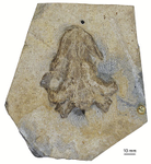
In the present study, Anquetin and his colleague Charlotte André extend the fossil record of these stem-turtles, recently hypothesized as a clade Thalassochelydia, into the Early Cretaceous (Anquetin & André 2020). They present an excellent anatomical account on a well-preserved cranium from the Purbeck Formation of Dorset (England) that can be referred to Thalassochelydia and augments our knowledge of the cranial morphology of this clade. Anquetin & André (2020) make a good case that this specimen belongs to the same taxon as shell material long ago described as Hylaeochelys belli.
References
Anquetin, J., & André, C. (2020). The last surviving Thalassochelydia—A new turtle cranium from the Early Cretaceous of the Purbeck Group (Dorset, UK). PaleorXiv, 7pa5c, version 3, peer-reviewed by PCI Paleo. doi: 10.31233/osf.io/7pa5c
The recommender in charge of the evaluation of the article and the reviewers declared that they have no conflict of interest (as defined in the code of conduct of PCI) with the authors or with the content of the article. The authors declared that they comply with the PCI rule of having no financial conflicts of interest in relation to the content of the article.
Evaluation round #1
DOI or URL of the preprint: 10.31233/osf.io/7pa5c
Author's Reply, 12 May 2020
Dear Editor,
Please find attached our revised article entitled "The last surviving Thalassochelydia—A new turtle cranium from the Early Cretaceous of the Purbeck Group (Dorset, UK)". We have carefully followed the suggestions of the two reviewers (see details below). All modifications are highlighted in yellow in the attached tracked changes pdf.
We have also slightly modified the Systematic paleontology part to include recent results from Evers & Joyce (2020) regarding the relative position of Thalassochelydia and Sandownidae (see page 6).
We hope that that you will consider this revised version to be of sufficient quality to be recommended by PCI Paleo.
Comments from Igor Danilov
Figure 4 (page 7): the reviewer requested some additional features to be labelled, notably the processus trochlearis oticum, incisura columellae auris, processus articularis of the quadrate, and infolding ridge on the quadrate. The processus trochlearis oticum (pto) was already labelled. We have added the infolding ridge on the quadrate (qr, for quadrate ridge). The other features are not apparent/preserved in the figure. The reviewer also suggested that we should add additional views (lateral, anterior, posterior) of the cranium. However, we feel that these views would not bring any meaningful value to the illustration because the specimen is severely flattened dorsoventrally. This is why we decided to refer the reader to the provided 3D model instead. This model provides a much better understanding of the complex morphology of the specimen at hand.
Note on the internal carotid arterial system in Paracryptodira: the reviewer suggested to add an illustration to compare the conception of previous authors to our own on this point. We feel this is unnecessary for two reasons. 1/ Our interpretation is actually not radically different from that of original authors. Instead, other authors have simplified the condition of the internal carotid arterial system in early paracryptodires to fit into exploitable phylogenetic characters. Therefore, we think that referring the reader to the original illustrations is enough. 2/ The concerned basal paracryptodires (notably Mesochelys/Pleurosternon and Uluops) are currently being redescribed based on 3D CT data by Walter Joyce and colleagues. These redescriptions will provide a much more reliable source of information than our few rapid notes resulting from the comparison of our new material to non-baenid paracryptodires based mostly on published literature.
The reviewer also asked that we mention that preliminary results of this study were presented at the 2018 Turtle Evolution Symposium in Tokyo and published subsequently as an extended abstract. We added a sentence to this purpose in the Introduction (line 35).
Comments from Serjoscha Evers
Carotid description of new skull: the reviewer asked that we provide more details about variation in thalassochelydians. We have added two sentences (see line 363) to talk about the variation in the group and refer the reader to two recent publications in which we provide de survey of this character.
Paracryptodiran carotid system: the reviewer wanted us to comment on the potential impact of our observations for paracryptodiran monophyly. We have added a sentence to the discussion (line 642) hitting toward the future results of Serjoscha's work, but we cannot really say more at the moment based only on our study.
Additional citations: most of the suggested references have been added.
Specific comments: we accepted the majority of the specific comments (see lines 90, 231, 265, 267, 294, 302, 315, 341, 375). We checked the position of the foramen supramaxillare, but it turns out we are now uncertain about our initial interpretation because the area is poorly preserved. We preferred to remove this mention altogether.
Sincerely yours,
Jérémy Anquetin and Charlotte André.
Decision by Hans-Dieter Sues, posted 26 Feb 2020
Both reviewers explicitly noted the scientific value of the contribution, and I concur. The authors should address the comments made by both reviewers but this should be easily accomplished.
Reviewed by Igor Danilov, 13 Feb 2020
This preprint is devoted to a detailed description of a new turtle cranium from the Early Cretaceous of the Purbeck Group (Dorset, UK), which is attributed to Thalassochelydia indet. This cranium may belong to the shell-based thalassochelydian species Hylaeochelys belli, known from the same deposits. The taxonomic attribution of the cranium is supported by a phylogenetic analysis based on one of the most recent global morphological matrix for turtles (Evers and Benson, 2019). In addition, to discussion of the beta and alpha taxonomy of the cranium, the preprint contains some notes on the internal carotid arterial system in Paracryptodira. The preprint is well and clearly written and I have only few comments and suggestions, which, I hope, will help to improve the preprint. 1) I suggest to add to figure 4 more designations of morphological structures described in the text, like the processus trochlearis oticum, the incisura collumelae auris, processus articularis of the quadrate, ridge on the posterior surface of this processus etc. 2) I suggest to add additional views of the specimen: right and left lateral, posterior and anterior. 3) An additional schematic illustration is desirable for the section "Notes on the internal carotid arterial system in Paracryptodira" to illustrate previous and present authors opinion about structure of this system in the turtles under discussion. 4) The authors should mention that preliminary results of this study have been published as Andre C. and Anquetin J. 2018. A new turtle cranium from the Early Cretaceous of the Purbeck Group (Dorset, UK). In: Hirayama et al. (Eds.). Turtle Evolution Symposium. Scidinge Hall Verlag Tübingen, ISBN 978-3-947020-06-5: 63-66.
https://doi.org/10.24072/pci.paleo.100008.rev11Reviewed by Serjoscha Evers, 24 Feb 2020
INTRODUCTORY NOTES
I enjoyed reading this well-written and well-illustrated MS and would like to congratulate the authors on their work. The descriptive work within this paper is comprehensive, and only restricted by the crushed preservation of the specimen. I appreciate the careful taxonomic approach of (i) not naming a new species on the basis of the new material based on the probability that the skull pertains to Hyaelochelys, but also (ii) being careful about the referral to Hyaelochelys, which is otherwise only known from postcranial material (i.e. there is no overlap in material that could provide concrete evidence for the referral). The authors perform a phylogenetic analysis, and they usage of the Evers & Benson (2019) matrix over alternatives is well reasoned and explained. Comparative descriptive comments as well their phylogeny support the systematic identification of their new material as belonging to the Thalassochelydia. Re-running their deposited matrix reveals the exact results as presented in this paper. The deposition of the 3D model of the cranium is much appreciated, facilitated easy review, and will be useful for future systematic work including this taxon. The comparative work aimed to distinguish the new specimen from paracryptodiran turtles reveals some interesting observations on the latter group. This extra bit of information makes the present paper relevant beyond thalassochelydian anatomy and systematics. I have a few specific comments, and also very few general comments that I would like to see addressed. All of those are outlined allow. However, all potential changes to the manuscript are very minor, so that I hope that this paper can be published in due course. With best regards, Serjoscha Evers
GENERAL COMMENTS
Carotid description of new skull In the descriptive section of the features related to the carotid circulation, it would be nice if the authors differentiate the patterns for thalassochelydians a bit more clearly. For instance, the ventrally exposed groove for the internal carotid artery is compared to other thalassochelydians by stating: “This is clearly reminiscent of the condition in some thalassochelydians, such as Plesiochelys etalloni, Plesiochelys bigleri, and Jurassichelon oleronensis (Gaffney, 1976; Rieppel, 1980; Anquetin et al., 2015; Püntener et al., 2017).” However, this list disregards variation among thalassochelydians: Although the arteries are indeed exposed in a trough in Plesiochelys bigleri and Jurassichelon oleronensis, the course of the internal carotid is ventrally entirely covered by bone in Portlandemys (Gaffney 1975) and Neusticemys (Gonzalez-Ruiz et al. 2019) [and also Solnhofia, although this taxon probably is a sandownids rather than a ‘true’ thalassochelydian], whereas it seems largely embedded in Plesiochelys planiceps (own observation; but see also works of Gaffney). The situation in Plesiochelys etalloni may be variable across specimens, but at least in NMS 40870, the artery lies in a trough anteriorly, but is posteriorly embedded, so that in this specimen, technically a fenestra caroticus (sensu Rabi et al. 2013) is present. I would recommend that the authors include these additional taxon/specimen citations, because only the full list gives an unbiased view on the variation that is present within thalassochelydians. Additionally, I would suggest to cite Raselli & Anquetin (2019) in this section, as that paper specifically compares at least some aspect of carotid variation among plesiochelyids.
Paracryptodiran carotid system I appreciate the comments of the authors regarding the paracryptodiran carotid system, because they align well with my own observations. The forward position of the fpcci (i.e. the posterior foramen for the internal carotid artery) has long been stated as basically the only synapomorphy of paracryptodires (i.e. some concept of baenids + pleurosternids). I agree with the authors, however, that the pleurosternids system is quite different from that of baenids, and that the proposed and often-cited synapomorphy is indeed not present. It would be interesting if the authors could comment on potential consequences of this observation. In my latest phylogenetic developments of the Evers & Benson (2019) matrix (not yet published), I fail to retrieve a monophyletic Paracryptodira, and upon checking the literature, there are actually very few global studies that do ‘manage to get’ a monophyletic Paracryptodira (and they all include the erroneous synapomorphy of the carotid system). Do the authors think that they’re carotid observations have any consequences for paracryptodiran relationships, and if so, how?
Additional citations I have suggested a few additional citations throughout my comments, but I want to highlight that I think the cranial description of Neustiqemys neuquina (Gonzalez Ruiz et al. 2019; Journal of Palaeontology, doi: 10.1017/jpa.2019.74); the paper discussing carotids in some specimens of Plesiochelys (Raselli & Anquetin. 2019. PLoS ONE 14(5): e0214629); one or several papers for protostegid comparisons regarding the laterally open foramen palatinum posterius (e.g. Williston 1898; Hirayama 1998; Kear & Lee 2006; Cadena & Parham 2015; Raselli 2018; Evers et al. 2019); and possibly the recent cranial description of Sandownia harrisi (Evers & Joyce. 2020. Royal Society Open Science 7: 191936) could be cited. For full disclosure, the Sandownia paper is a recent paper of mine, and the authors could not have known of this paper when the prepared the MS at hand. So I consider this an optional additional citation.
SPECIFIC COMMENTS In line 96, the authors write “For a recent reassessment of basal paracryptodire taxonomy, the reader is referred 96 to Joyce and Anquetin (2019).“ I disagree with the use of the word ‘basal’ here: The primary comparisons listed in this section are Glyptops and Pleurosternon, both members of the Pleurosternidae. Pleurosternids are most commonly inferred to be the sister-group to baenids. However, as the sister to baenids they are no more ‘basal’ than baenids themselves. I think this problem can be easily circumvented by using ‘non-baenid paracryptodire’ or ‘pleurosternid’ instead of ‘basal paracryptodire’. Line 243ff: Regarding the jugal description, the authors do not comment about whether a medial jugular process is present or absent. This process is commonly absent in plesiochelyids or thalassochelydians, and it seems that this is also the case here, although the region is obscured by crushing… It would be nice if the authors could specifically comment on this feature and its preservation. Line 280/281: The usage of ‘arms’ in respect to the diverging rami of the maxillae is somewhat unusual, and I recommend rephrasing the sentence. Line 282: The authors describe the lingual ridge of the maxilla to be ‘serrated’. I have checked the images and the 3D file, and know what is meant. However, the pattern is a bit irregular on both skull sides, and serrations immediately remind of testudinid-like structures that are present in the rhamphotheca. Do the authors think that the observed ‘serrations’ are features that would have been mirrored in the horny beak, or are those simply roughened areas of bone, possibly linked to the innervation and blood supply of the beak? It would be nice to comment on this with one or two sentences. In line 306, the authors note that “The foramen supramaxillare opens in the posterior part of the orbit floor along the suture between the maxilla and jugal (visible only on the left side)”. This is interesting, because we recently described this foramen to be within the jugal of Sandownia harrisi (also an angolachelonian; Evers & Joyce 2020). The foramen supramaxillare is usually fully and clearly formed by the maxilla, so the position of this foramen here in comparison with Sandownia is intriguing. I leave it open if the authors want to include a comparison with Sandownia, as it is a paper of my own, and as it only came out so recently that the authors could not have included this particular comparison a few weeks ago. line 310: ‘excepting’ seems to be linguistically incorrect in this instance. I would suggest using ‘with exception of’ instead. Line 315 ff: Palatine description. The presence/absence of an interpalatine/vomer-pterygoid contact is only briefly mentioned in the section describing the pterygoid. The authors seem to infer that an interpalatine contact is absent. This has also been described for many thalassochelydians (e.g. Neusticemys; Gonzalez-Ruiz et al. 2019; see also Gaffney 1975), but not for all (e.g. Plesiochelys planiceps: Gaffney 1975). Given that the respective sutures are difficult to see in the specimen at hand, it would be good of the authors discuss this particularly possible contact, or their interpretation thereof, in a bit more detail here, possibly citing the above studies that provide evidence for variation of this feature in thalassochelydians. In line 327, the authors write “It is known only in some thalassochelydians (Plesiochelys spp. and Jurassichelon oleronensis) and some early pan-chelonioids, whereas modern sea turtles lack the foramen altogether (e.g., Gaffney, 1976; Joyce, 2007; Anquetin et al., 2017)”. I think it would be helpful to cite instances of pan-chelonioids that have the feature of the ‘open’ foramen palatinum posterius. Citing instances would be important, because the feature is not present in ‘random’ pan-chelonioids, but in protostegids (e.g. Williston 1898; Hirayama 1998; Kear & Lee 2006; Cadena & Parham 2015; Raselli 2018; Evers et al. 2019). Protostegids have repeatedly been hypothesized to be closely related to thalassochelydians (e.g. Joyce 2007; but many others), so that this comparison is highly relevant to both the positions of thalassochelydians and protostegids – both of which are debated. Line 351: The infolding ridge of the quadrate is also present in sandownids, and not only in thalassochelydians (Evers & Benson 2019; Evers & Joyce 2020). Line 383: ‘briefly’ is usually a term used for durations. I would recommend something like ‘along a short contact’ line 544: the word ‘sterile’ seems misplaced in this context; maybe exchange with something like ‘acertained’? line 568: you could delete the ‘somewhat’; the conclusion is definitely supported by your results. line 568: Please include a figure reference to Fig 6. here.
Download the review https://doi.org/10.24072/pci.paleo.100008.rev12


