
Inferences on the lifestyle of fossil xenarthrans based on limb long bone inner structure
Palaeobiological inferences based on long bone epiphyseal and diaphyseal structure - the forelimb of xenarthrans (Mammalia)
Abstract
Recommendation: posted 19 September 2018, validated 22 September 2018
Houssaye, A. (2018) Inferences on the lifestyle of fossil xenarthrans based on limb long bone inner structure. Peer Community in Paleontology, 100001. https://doi.org/10.24072/pci.paleo.100001
Recommendation
Bone inner structure bears a strong functional signal and can be used in paleontology to make inferences about the ecology of fossil forms. The increasing use of microtomography enables to analyze both cortical and trabecular features in three dimensions, and thus in long bones to investigate the diaphyseal and epiphyseal structures. Moreover, this can now be done through quantitative, and not only qualitative analyses. Studies focusing on the diaphyseal inner structure (cortical bone and sometimes also spongious bone) of long bones are rather numerous, but essentially based on 2D sections. It is only recently that analyses of the whole diaphyseal structure have been investigated. Studies on the trabecular architecture are much rarer.
Amson & Nyakatura (2018) propose a comparative quantitative analysis combining parameters of the epiphyseal trabecular architecture and of the diaphyseal structure, using phylogenetically informed discriminant analyses, and with the aim of inferring the lifestyle of extinct taxa. The group of interest is xenarthrans, one of the four major extant clades of placental mammals. Xenarthrans exhibit different lifestyles, from fully terrestrial to arboreal, and show various degrees of fossoriality. The authors analyzed forelimb long bones of some fossil sloths and made comparisons with several species of extant xenarthrans. The aim was notably to discuss the degree of arboreality and fossoriality of these fossil forms.
This study is among the first ones to conjointly analyze both diaphyseal and trabecular parameters to characterize lifestyles, and the first one outside of primates. No fossil form could undoubtedly be assigned to one lifestyle exhibited by extant xenarthrans, though some previous ecological hypotheses could be corroborated. This study also raised some technical challenges, linked to the sample and to the parameters studied, and thus constitutes a great step, from which to go further.
References
Amson, E., & Nyakatura, J. A. (2018). Palaeobiological inferences based on long bone epiphyseal and diaphyseal structure - the forelimb of xenarthrans (Mammalia). bioRxiv, 318121, ver. 5 peer-reviewed and recommended by PCI Paleo. doi: 10.1101/318121

The recommender in charge of the evaluation of the article and the reviewers declared that they have no conflict of interest (as defined in the code of conduct of PCI) with the authors or with the content of the article. The authors declared that they comply with the PCI rule of having no financial conflicts of interest in relation to the content of the article.
Evaluation round #2
DOI or URL of the preprint: 10.1101/318121
Version of the preprint: 2
Author's Reply, 11 Sep 2018
Decision by Alexandra Houssaye, posted 11 Sep 2018
Dear authors,
Thank you for submitting a revised version of your manuscript entitled “Palaeobiological inferences based on long bone epiphyseal and diaphyseal structure - the forelimb of xenarthrans (Mammalia)“. You made significant changes to your manuscript, in order to address the points raised by the reviewers. I checked the whole manuscript and I have only a few comments that I wrote directly in the manuscript before to recommend it. I will send it to you by email because we can not send it via the website yet. Could you please consider the remarks and submit a new version accordingly. Best wishes,
Alexandra Houssaye
Evaluation round #1
DOI or URL of the preprint: 10.1101/318121
Version of the preprint: 1
Author's Reply, 04 Sep 2018
Decision by Alexandra Houssaye, posted 04 Sep 2018
Dear authors,
I have agreed to handle the evaluation of a preprint entitled "Palaeobiological inferences based on long bone epiphyseal and diaphyseal structure - the forelimb of xenarthrans (Mammalia)", for potential recommendation by Peer Community in Paleontology (PCI Paleontology).
The preprint was sent to two reviewers. If they both highlight the interest and quality of the preprint, they also suggest moderate/major revision. Their comments, which should enable you to greatly improve the preprint, are attached. Below, you will also find additional comments provided by a PCI Paleo recommender.
Thank you again for submitting your interesting work to PCI Paleo. I look forward to receive your revision.
Best wishes,
Alexandra Houssaye
Additional comments from a PCI Paleo recommender:
- Suggestion to use Folivora rather than Tardigrada
- l. 111. Suggestion to cite Pujos et al. 2012 Figure 4 Pujos, F., Gaudin, T. J., De Iuliis, G., & Cartelle, C. (2012). Recent advances on variability, morpho-functional adaptations, dental terminology, and evolution of sloths. Journal of Mammalian Evolution, 19(3), 159-169.
- l. 236. Suggestion to compare with the two last molecular publications for the age of the split between Bradypus & Choloepus (Slater et al. 2016; Delsuc et al. 2018) Slater, G. J., Cui, P., Forasiepi, A. M., Lenz, D., Tsangaras, K., Voirin, B., ... & Greenwood, A. D. (2016). Evolutionary relationships among extinct and extant sloths: the evidence of mitogenomes and retroviruses. Genome biology and evolution, 8(3), 607-621. Delsuc, F., Philippe, H., Tsagkogeorga, G., Simion, P., Tilak, M. K., Turon, X., ... & Douzery, E. J. (2018). A phylogenomic framework and timescale for comparative studies of tunicates. BMC biology, 16(1), 39. -l.419. Considering the strong uncertainties relative to the position of Hapalops (notably linked to strong changes in sloth phylogeny with the new results coming from aDNA), you should say on world on this as well, to moderate your results based on phylogenetically informed analysis. If you change the position of Hapalops (for example closer to Bradypus), dies it change the result?
- l. 462. You cite a manuscript under review.
- l. 489. It is not only argued that Xenarthra would be one of the major clades of placental mammals; it is one of the 4 early diverging clades of extant placentals; you might cite molecular analyses for that as well.
Reviewed by anonymous reviewer 1, 19 Jul 2018
The manuscript of Amson and Nyakatura is an interesting contribution to the existing literature on the relationship between trabecular and cortical bone and behaviour of extant xenarthrans, and tests whether the behaviour of fossil taxa can be reconstructed. In their previous publications, the authors have been the first researchers to apply modern methods to investigate trabecular bone structure in non-primate taxa, and I am pleased to see the potential utility of this aspect of bone structure explored.
The humerus and radius are analysed amongst extant taxa which differ in both their locomotor mode, and in the frequency and mode of fossorial behaviour. Several fossil xenarthrans were analysed in order to determine their locomotor and fossorial behaviour. Although, locomotor and fossorial behaviour could not be not confidently assigned to the fossil taxa included in the analysis, Hapalops was found to have sloth-like morphology. I am pleased to see the authors discuss the complexities of this methodology, including the problems arising when interpreting bone structure in such large-bodied, extinct species.
I have divided my comments on the manuscript into major and minor suggestions, and have also noted spelling and grammatical errors.
Major comments
1. The manuscript would greatly benefit from a more detailed and nuanced interpretation of the various bone parameters measured.
Cortical and trabecular bone are analysed collectively in the manuscript, however, the two regions of bone perform very different functions. Cortical bone cross sectional geometric properties reveal the potential for bones to resist compressive and bending forces, whereas trabecular bone structure is adapted to compressive forces at the articular surface. As such, the two types of bone would not necessarily be expected to co-vary directly, as assumed by the authors.
The manuscript should more clearly explain how these two different bone structures reflect mechanical loading.
2. A separate, but related comment is that the biomechanical hypotheses tested are not clearly explained. I understand that often the kinetics/kinematics of behaviours such as climbing and digging are unknown, but the authors should elaborate on the differences that would be expected for their behavioural categories. For example, what differences might there be in the variability of loading (as measured by DA) between fossorial and non-fossorial groups? Or, how might the overall the magnitude or orientation of loading differ between behavioural categories?
Although not feasible to incorporate in the present study, I wonder if elements of the hindlimb might be informative for future studies. The two behaviours of interest in these groups are arboreal/terrestrial locomotion and fossorial behaviour. I assume that digging in these groups only, or at least primarily, involves the forelimb, in which case you would expect the hindlimb to be influenced only by locomotion. As it stands, the elements in this study are influenced by both locomotion and digging, thus the morphology is likely due to a combination of two different loading regimes.
3. The cross-sectional geometry of the elements included in this study are highly complex, and at the sites analysed there are large muscle attachment sites. I would suggest that the authors discuss the possible impact of these muscle attachments sites on the cross-sectional geometry results, and for future analyses consider sampling locations on the diaphysis without prominent entheses.
4. Information should be included on sample size and extant species in the sample, and the details of CT scanning of the extant sample (i.e. where they were scanned and at what resolution).
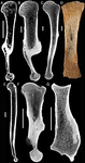
5. I would like to see more information about the VOI placement protocol. The methods say that ROIs were selected from the centre of the epiphyses, however, looking at the images in Amson et al. 2017, this doesn't appear to be the case for either the humeral head or the radial trochlea, the MC3 was not included in this previous publication. A clearer description, and preferably a figure, should be included to explain further the VOI placement protocol.
6. p10, line 223: Was the total volume used here the size of the VOI? This is unlikely to be a good size proxy, because the VOIs were not scaled to the size of the epiphysis, rather as large a VOI as possible, avoiding cortical bone, was placed in the epiphysis. Although the TV is not used as a size proxy in the analysis, a measure of the size of the epiphysis would be more appropriate than the size of the VOI.
7. p12, line 265: Were the parameters normally distributed after log-transformation?
8. The paper should include a results table with the mean values for each taxonomic group or species, and the results for each fossil.
9. p13, Univariate Comparisons: The focus of this section is on the fossil taxa, but it would help the reader a brief description of how the extant groups differ from one another was included, for all parameters discussed.
10. Not enough information is given concerning the discriminant function analysis -- I would expect the paper to include a table reporting data from this analysis, and additional information in the text. For example, a table with predicted group membership should be included for both the training data and the fossil taxa. It would also be informative to include the contribution of the variables to each function to better understand which variables are driving between group differences. How were the extant taxa grouped -- it is unclear whether this is at the species level, generic level, or by a behavioural classification?
11. What is the potential influence of correlation between variables on the DFA, and on the PCA used for Hapalops? The included trabecular parameters are likely to be correlated with one another, for example BV/TV and Tb.Th.
Minor comments
Abstract: The authors overstate the sensitivity of trabecular architecture by using the phrase "extreme accuracy and sensitivity" in the abstract; in p1 line 44 "great accuracy and sensitivity"; and in p1 line 47 "great plasticity". Studies in primates have had very mixed results, in many cases the relationship between trabecular structure and behaviour is unclear. I would recommend these phrases are adjusted to reflect that it is not known how accurately trabecular structure reflects loading.
P4, line 65: Tsegai et al. (2017) used the cortical thickness method developed by Treece et al. (2010; 2012). It is important to note that the focus was cortical bone thickness at the articular surface, rather than diaphyseal structure.
p4, line 88 and p7, line 153. Anteaters are described here as intermediate, it should be more specific, is this intermediate in their fossorialism or terrestrial/arborealism?
p9, line 209: Change "trabecular" to "cortical"
p17, line 401: Include other publications from the primate literature, as there any many studies which find DA, or primary trabecular orientation, to be informative (e.g. Ryan and Ketcham, 2002; Griffin et al., 2010; Barak et al., 2013; Su et al., 2013).
p19, line 460: This is in important point, which could be expanded upon. Is there any evidence for this in extant xenarthrans?
Spelling/grammatical comments:
p3, line 44: "excepted" should read "expected"
p3, line 49-54: Using "was" here sounds rather strange, I would recommend using "has been". Also on p4 line 73, "parameters were" should read "parameters have never been", also p18 line 430 change "was never" to "has never been".
P4, line 68: Here and in a few other instances throughout the manuscript there is an error in the number of parentheses.
p5, line 91: "this is likely not true anymore" is confusing -- please rephrase.
p5, line 95: Change teeth to tooth. It is either tooth morphology or morphology of teeth. p5, line 97: In "This was found as challenging" change "as" to "to be".
p5 line 101: Remove first "in"
p5 line 113: Add "a": "Such a lifestyle".
p10, line 224: Change "specific" to "species".
p10, line 228: Add "taxa" after "extinct".
p11, line 245: Change "Beside" to "Besides"
p12, line 270-271: There is an error with the ü in the PDF.
p12, line 274: Indent subheading
p16, line 383: Change "correlated to" to "correlated with"
p16, line 384: Change "studied taxa to influence the analysis" to "studied taxa from influencing the analysis"
p18, line 432: Change "as consistent with a fossorial" to "to be consistent with fossorial" p18, line 434: Change "non-fossorial taxa to" to "non-fossorial taxa in"
p18, line 439: Move "is", this should read "in neither case is the classification clear"
p 18, line 440: Change "more of their" to "additional"
p21, line 488: Change "investigations" to "investigation"
References
Barak MM, Lieberman DE, Raichlen D, Pontzer H, Warrener AG, Hublin J-J (2013) Trabecular evidence for a human-like gait in Australopithecus africanus. PLoS ONE, 8, e77687.
Griffin NL, D'Août K, Ryan TM, Richmond BG, Ketcham RA, Postnov A (2010) Comparative forefoot trabecular bone architecture in extant hominids. J Hum Evol, 59, 202-213.
Ryan TM, Ketcham RA (2002) The three-dimensional structure of trabecular bone in the femoral head of strepsirrhine primates. J Hum Evol, 43, 1-26.
Su A, Wallace IJ, Nakatsukasa M (2013) Trabecular bone anisotropy and orientation in an Early Pleistocene hominin talus from East Turkana, Kenya. J Hum Evol, 64, 667-677.
Treece GM, Gee AH, Mayhew PM, Poole KES (2010) High resolution cortical bone thickness measurement from clinical CT data. Medical Image Analysis, 14, 276-290.
Treece GM, Poole KES, Gee AH (2012) Imaging the femoral cortex: Thickness, density and mass from clinical CT. Medical Image Analysis, 16, 952-965.
Download the review https://doi.org/10.24072/pci.paleo.100003.rev11Reviewed by Andrew Pitsillides, 21 Jul 2018
Overall comments: This work in this manuscript seeks to establish relationships between known lifestyles and cortical and trabecular parameters, in a range of bones of extant animals from a specific clade. The intention seems to be, to use any relationship that emerges to make predictions regarding the lifestyles in extinct clade members, based solely upon scans of their fossilised bone remnants. The authors present a combined analysis of cortical and trabecular parameters of the xenarthran forelimb, based on data from one of their group’s previous publications (trabecular bone of extant xenarthrans) supplemented with novel trabecular data from extinct tardigrades (ground sloths) and cortical data from all xenarthrans. Whilst the previous study, predictably found that forelimb function matched the phylogeny – thus, that “all armadillos dig, all sloths climb and all anteaters do a bit of both” – this new manuscript, in contrast, finds instead that ground sloths don’t match the lifestyle of the extant members of their clade. Major comments 1. The authors claim to have identified the ‘challenges’ to making any such predictions with security. These are stated to be: i) the imperfect lifestyle discrimination for the extant animals, ii) the difficulties of scaling of these parameters for extinct animals, which are outside the range for extent animals and iii) the classification of the values from extinct animals as outliers with respect to the extent animals.
This reviewer is not expert enough in this area to know whether these ‘challenges’ could have been predicted before undertaking these analysis, nor whether their identification alone is novel enough to warrant reporting. Both need to be addressed by the authors and then an expert palaeontologist should verify the robustness of their response.
This applies particularly to their conclusions (and throughout); for example (line 120-123), where it was unclear to me what the purpose of this study was, unless it was to unambiguously identify the reasons that the reconstruction [lifestyle] of the extinct Xenarthrans cannot be achieved by the means they have used?
- The authors state (line 43) that trabecular bone reacts to loading very sensitively, which indeed it does rather rapidly too, to provide a ‘functional signal’. Does this bring into question the use of its analysis as a readout of likely ‘lifestyle’ in extinct animals? These extinct animals would have to have died in perfect ‘health’ in order that direct comparison with extant, presumably healthy, animals is to be made. This might indeed be the case and so the likely reason for their extinction is rather important and should be stated if known.
This point also raises another major worry; might this statement be deeply misleading. I would recommend the authors make it clear, that, although the fact that bone is typically responsive to loads, which parts of the bone respond to what extent is still very much unclear. As the review of trabecular bone functional adaptation cited by the authors (Kivell, 2016) states: “bone functional adaptation is not sufficient (but is all we have)”. I have no problems with the authors trying to infer lifestyle from the information they have, but they should make it clear that this approach is (necessarily) limited. Minor comments 1. Abstract: The last sentence is not clear and should be re-written. I think an issue with the phrasing is due to the use of the word ‘challenges’. Introduction: 2. I would like the authors to make the aim of the paper clear in the last paragraph of the introduction. Are they trying to reconstruct the lifestyle of extinct xenarthrans based on a combination of cortical and trabecular parameters (as I was led to believe until line 111), are they presenting a new method to reconstruct function from trabecular and cortical parameters (as suggested by the review of previous similar methods in the paragraph starting on line 55) or are they identifying challenges related to reconstructing lifestyle from trabecular and cortical parameters (as they state at the end of the introduction)?
This issue occurs elsewhere and my preference would be for this to be revisited (throughout) to add greater meaning to the conclusions. Current phrasing suggests that the authors have set out to identify whether there are ‘challenges’ rather than address questions that could realistically have been addressed using the samples available.
- Line 12: “relative number” relative to what?
- Line 45. accepted for ‘excepted’
- Line 66: “same” it is not clear to me whether this refers to the current study or to Gross et al
- Line 70: “distinct zones of different arrangement” I presume this refers to e.g. what is sometimes called the vertical and horizontal trabecular columns in the femoral neck (Hammer, 2010, Annals of Anatomy, “The structure of the femoral neck: A physical dissection with emphasis on the internal trabecular system”) but this is not clear… could the authors please clarify?
- Line 79: I do not understand why a medulla full of trabecular bone helps withstand compression. Could the authors elaborate?
- Line 85: Could the authors please expand on the findings of their previous paper (Amson et al, 2017a) here? I would help set the context for the next paragraph. Page 5, line 89-92: it is not clear to this reader exactly what point is being made here. Suggest rephrasing.
- Line 104: I presume the phrases/terms autapomorphic nature, phylogenetic signal and ecophenotypic character are used appropriately? How can “ecophenotypic character” be a “rationale”? And what is an ecophenotypic character? Please clarify.
- Line 91: I understand what the authors mean by “likely not true anymore”, but this should be clarified, e.g. by adding a clause about why this is “not true anymore” at the end of this sentence.
- Line 119: It seems to me that the large-sized ground sloths exceed extant xenarthrans by more than an order of magnitude in body mass? Materials and methods
- The exclusion criteria on page 8 are not readily understood. In general, the methods are very discursive. I am not sure whether this is acceptable or not. Might it be more meaningful to move some of this apparent ‘validation/deliberation’ (pages 10-11) to the discussion?
- It would be good practice if the authors provided their R-code online as supplementary material, it may be useful for others to replicate their results/use their method on a different data set.
- Line 146: I believe this is a typo: “scanning resolution ranged from 0.03 to 0.123 micrometer” - shouldn’t this be 0.123 millimeter or 123 micrometer? To my knowledge, typical microCT scans performed in our lab on mouse tibiae result in a pixel size of several micrometers to several tens of micrometers – 0.03 micrometer=30 nanometer which I don’t think is realistically possible in a reasonable amount of scanning time (especially for large species such as ground sloths) or necessary to gather trabecular geometry information?
- Line 203: I believe this is a typo: did the authors forget to abbreviate global compactness to GC in this sentence? Results
- Could the results be shortened?
- Univariate comparisons: It is unclear to me how the authors choose the anatomical locations (% length) to compare the cortical parameters in humerus and radius to each other: sometimes it’s mid-diaphysis, sometimes 72% and sometimes 35%. I assume Figure 3 shows mid-diaphyseal data? This should be made clearer. If the authors compare data from different anatomical locations to each other, then I would appreciate a sentence on their thoughts about how valid these comparisons are.
- Lines 342, 348, 354: I believe this is a typo: this should be “could be included”, not “could have been included”? Discussion
- Page 16, lines 380-382: Has this been shown previously? If so, then requires a reference.
- Page 19: The most dramatic factors affecting bone structure during the life-course are likely age, gender and health status. This needs to be dicussed.
- Lines 389-397: I don’t understand the rationale behind proposing a size-corrected DA – it is a dimensionless parameter. Also I find the statement that DA scaled negatively in Doube et al, 2011 misleading: since (as the authors state) the p-value was >0.05, surely one would conclude that DA does not scale? Concluding remarks
- I find the paper an interesting follow-up on the authors’ group previous work on the xenarthran forelimb microarchitecture. The authors present some interesting thoughts on reconstructing anatomical function from as much information as is available in a clade that is not typically studied. I feel strongly that the authors should explicitly make clear that although bone responds to load, the nature of this response is a lot less predictable than the authors make it out to be currently. I feel the paper would further benefit from several clarifications as well as a more clear overall storyline to guide the reader through the ideas.


